Pulsed radiofrequency treatment to the occipital nerve can help to ease the pain of this condition, and associated scalp pain and sensitivity. It is a minimally invasive procedure which can be repeated to maximise the pain relieving benefits.
Why do this procedure? (Indications)
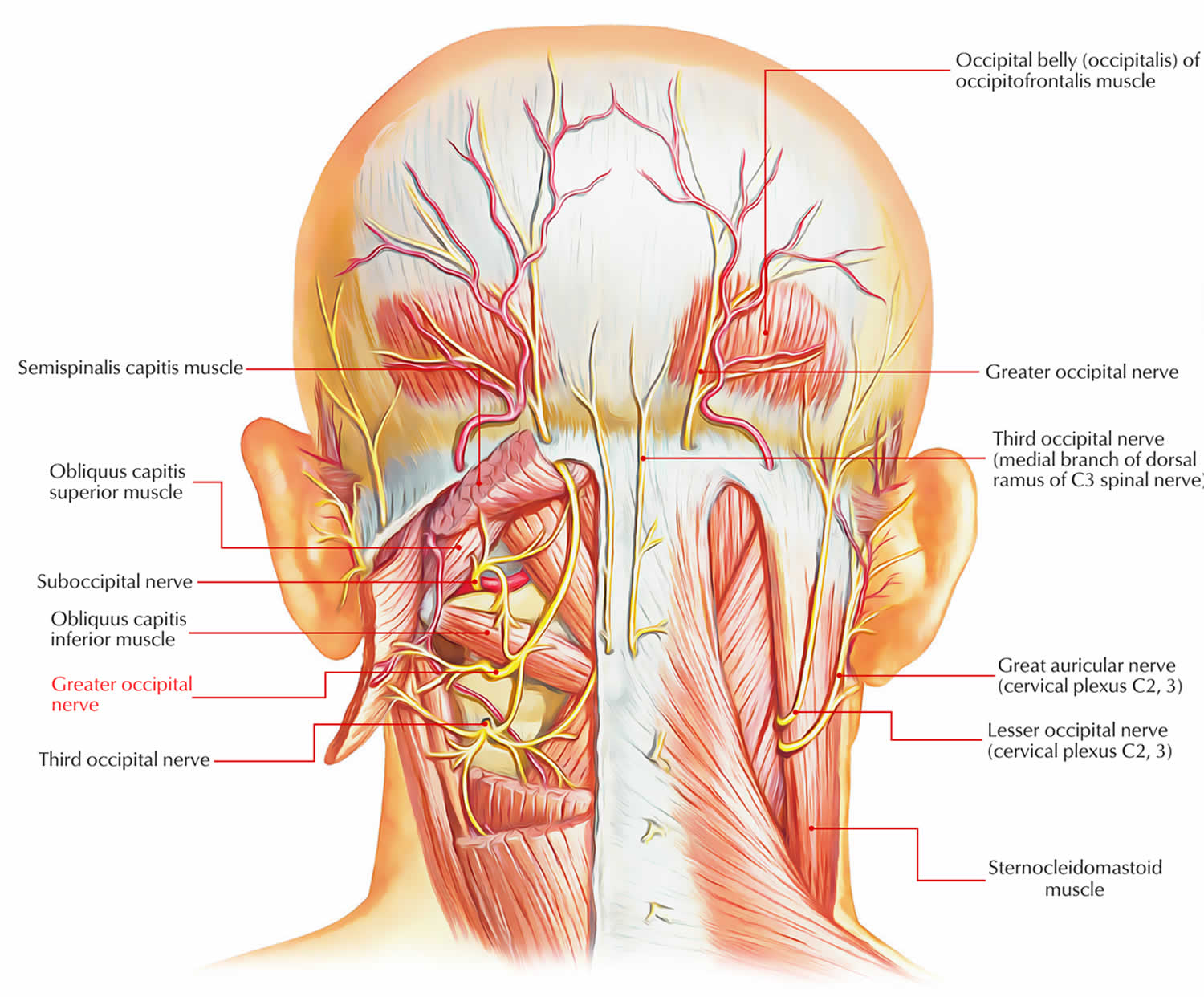
Pain felt around the occipital (back of head) region is often transmitted to the brain via these Occipital Nerves. Patients suffering from this type of pain are good candidates for an injection to the Occipital Nerves in order to disrupt the pain signals reaching the brain. If the injection contains a long acting steroid such as Betamethasone (Celestone) then patients may get long-term relief.
If the patient does have relief from their pain after the injection has been performed but their pain returns at some stage then there are 2 options
- Repeat the injection – especially if the patient obtained long term relief; or
- Use the injection as the indication for Pulsed Radiofrequency to the Occipital Nerve sto possibly obtain longer term relief (this is a longer term treatment option)
What is Pulsed Radiofrequency to the Occipital Nerves?
The procedure can be performed under X-Ray Guidance (Image Intensifier), Ultrasound guidance or use patient sensory testing. This involves the patient identifying when the needle is close to the needle allowing the procedure to be effective. It is a sterile procedure, which passes a needle through the skin along the bottom of the skull between the midline and the ear (Mastoid Process) Local anaesthetic is usually injected under the skin before the needle is inserted. Once the correct position has been confirmed an electric pulse is applied to the Greater and Lesser Occipital Nerve in order to inhibit the transmission of pain signals.

If the procedure is successful; there is no way of knowing how long it will last. The Third Occipital Nerve is treated at a different site. It runs close to the outside of the Second and Third Cervical transverse process. Two needles are placed next to these sites and Radiofrequency neurotomy is used to treat the nerve
How effective will it be?
It is not possible to predict or guarantee the effectiveness of any treatment. However, the fact that this procedure has been suggested means that it may be of use.
There are a number of possible outcomes:
- It may treat the pain and the pain does not return
- It may treat the pain for a short period of time (weeks to months) and then the pain returns either less or the same as before. In this case a repeat procedure could be indicated.
- There may be no effect on the pain (failure to confirm the source of the pain)
How is the treatment performed?
The procedure will be done as a clinic or rarely as a day patient. The injections may be performed in day surgery using a mobile X-ray machine for guidance, or “blind” in clinic using anatomical landmarks. In theatre, you will be placed onto the procedure table face down, lying on some pillows for your comfort. We need to get you into the correct position before we start the procedure. As this procedure is usually done under sedation, you will need to starve yourself beforehand, from midnight the night before or morning after a light breakfast if your procedure is in the afternoon or as directed. You may take all your usual painkillers and other medication unless specifically advised. If you are on blood thinning agents (warfarin), please let the doctor know well in advance as special arrangements will have to be made. Please also bring a list of your medications and any allergies. You will need someone to drive you home after the procedure.

Procedure technique
Before we start the procedure in the operating theatre, a small intravenous cannula will be placed into a vein in your hand. This is routine and allows us to administer any medication including medication for sedation or fluids if necessary. Your lower back will be uncovered and cleaned with an antiseptic solution to prevent infection. Local anaesthetic is then used to numb the skin before starting. Further anaesthetic will then be used to numb the space between the skull and the skin in the region of the Occipital Nerves. A needle is guided into the correct area. Once in the space, an electrical current is applied and the patient feels a buzzing sensation that confirms the correct positioning. If this isn’t present the needle may need to be repositioned to the correct site.. If you are awake, sometimes a feeling of pressure develops in the skull and neck region – this is usually only mild and temporary. After this step you will be more heavily sedated to undergo the Radiofrequency neurotomy at C2 and C3 levels.

COMPLICATIONS
Common Complications
- Continuing pain / no benefit
- Minor bleeding in the area treated
- Bruising Numbness in the area treated
- Temporary weakness or numbness from the local anaesthetic
- Brief increased pain that may fluctuate in intensity
More Serious Side Effects
- Damage to surrounding structures
- Infection
- Permanent nerve injury
- Allergy to the anaesthetic drugs used as part of the procedure
- Increase of any pre-existing medical condition such as cardiac conditions
- Burning sensation / Bruising around the area from the procedure
- Aspiration during sedation
- Eye injury while lying prone (face down)
- Serious anaesthetic / procedural complications and very rarely stroke or death
- Increased lifetime risk of cancer due to X-rays exposure
- Very rare risk of surgery due too injuries from the procedure
Please discuss with your doctor any other questions you may have about this procedure or this information sheet. If you agree to have the procedure, you will be asked to sign a consent form.
If you notice –
- Any swelling from the site,
- Any bleeding from the site, or
- Have any other concern
Please contact your General Practitioner, Queensland Pain Clinic, or the Emergency Department of your local hospital.


